
Floraform – an exploration of differential growth
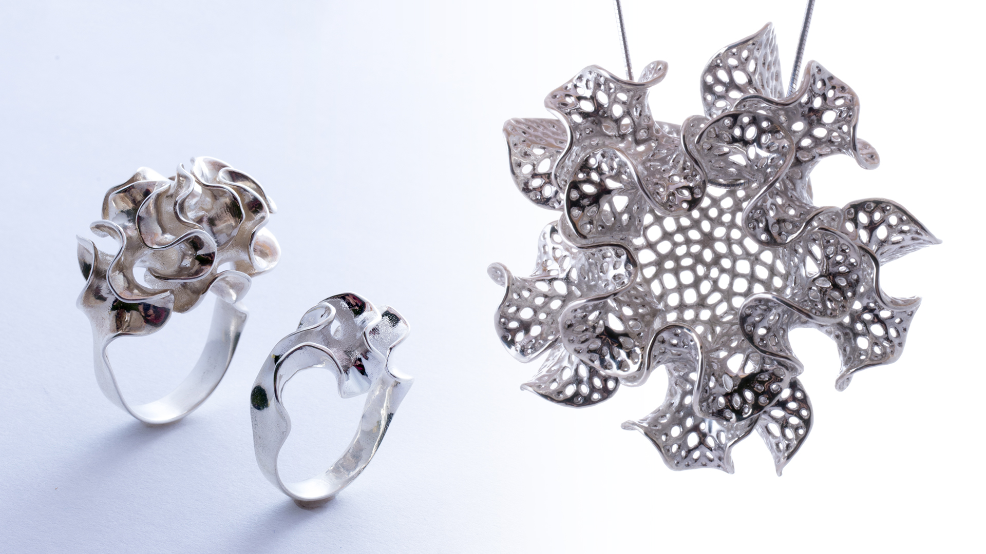
Introducing Floraform, the latest generative design system from Nervous System. Floraform is inspired by the biomechanics of growing leaves and blooming flowers and…
Introducing Floraform, the latest generative design system from Nervous System. Floraform is inspired by the biomechanics of growing leaves and blooming flowers and…
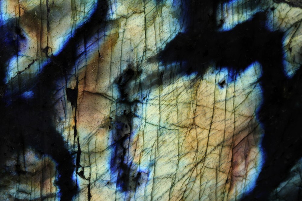
Labradorite is probably my favorite mineral. Ignore for the moment that it’s stunningly beautiful, and check out the killer science: Labradorescence The distinctive…
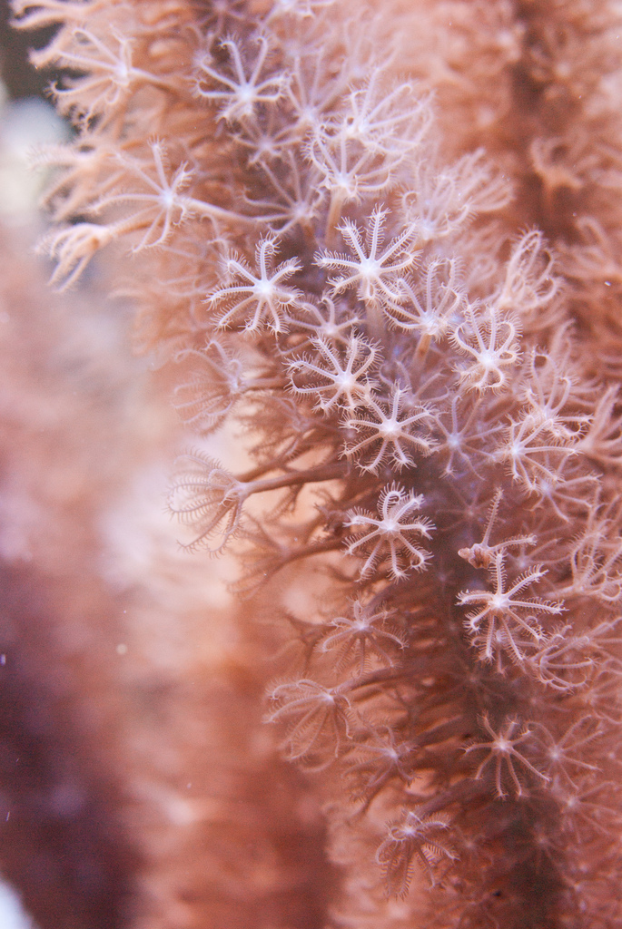
I’ve always been fascinated by coral. At first, for the otherworldly environments they create on the ocean floor. And later, for a whole…
Glyptodons are the extinct ancestors of modern day armadillos. These giant mammals roamed the Americas from 2.5 million years ago until just as…
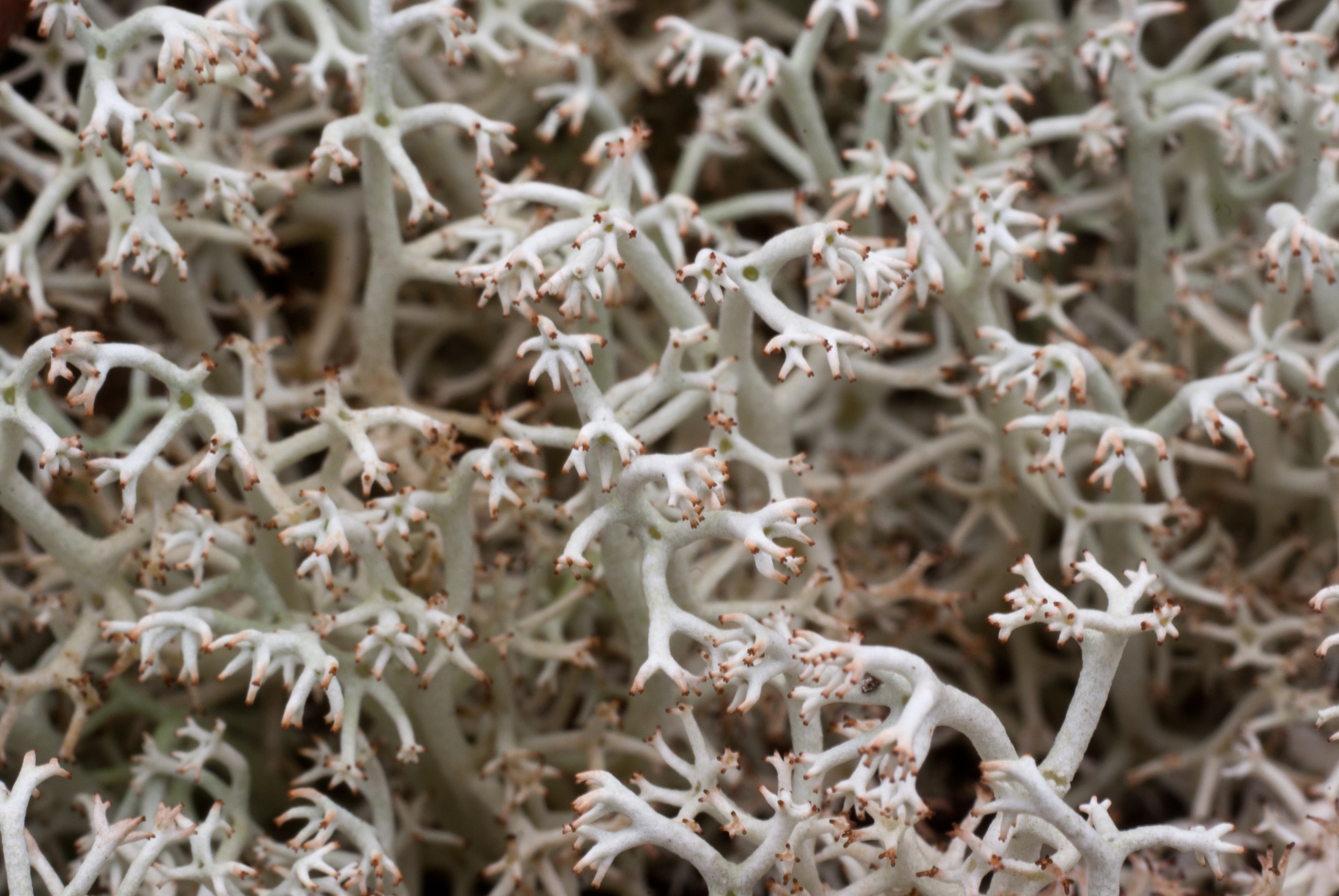
Recently, my friend Shaunalynn Duffy asked me to give a talk at a Sprout event centered around Fungi. Specifically she was interested in…
On Sunday, we visited the New England Aquarium in Boston, one of my favorite places in the city. Here are some photos I…
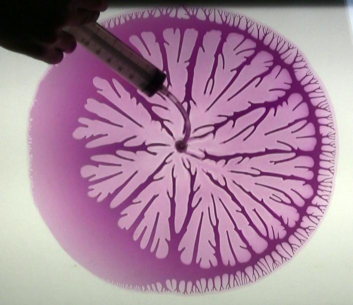
The video documents several Hele-Shaw Cell experiments using two 16×20″ panes of glass. Intricate branching patterns emerge as we insert glycerin, air and…
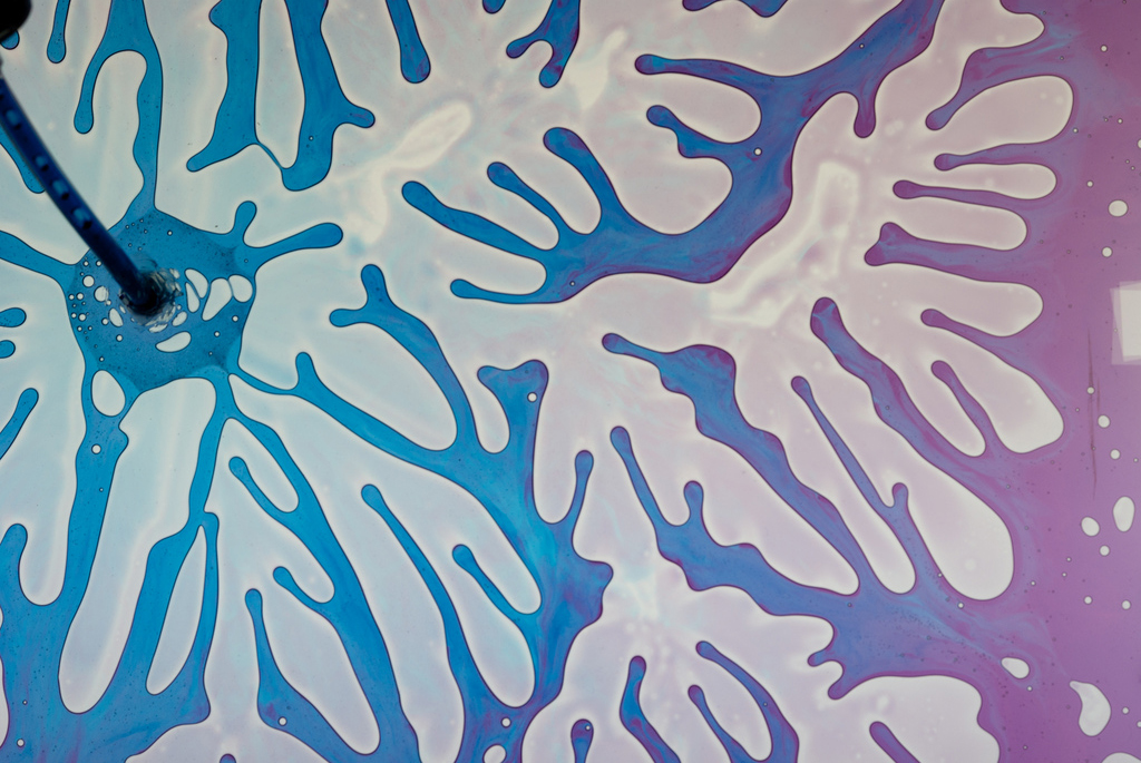
We spent Wednesday evening at Sprout working on a larger scale Hele-Shaw cell experiment and trying to figure out what materials and setup…
Nigel Goldenfeld is a professor of Physics at the University of Illinois, Urbana-Champaign who has worked on a wide-variety of fascinating topics. We…
When we start working on a new project, we do a lot of preliminary research. This usually involves a wide literature search on…