
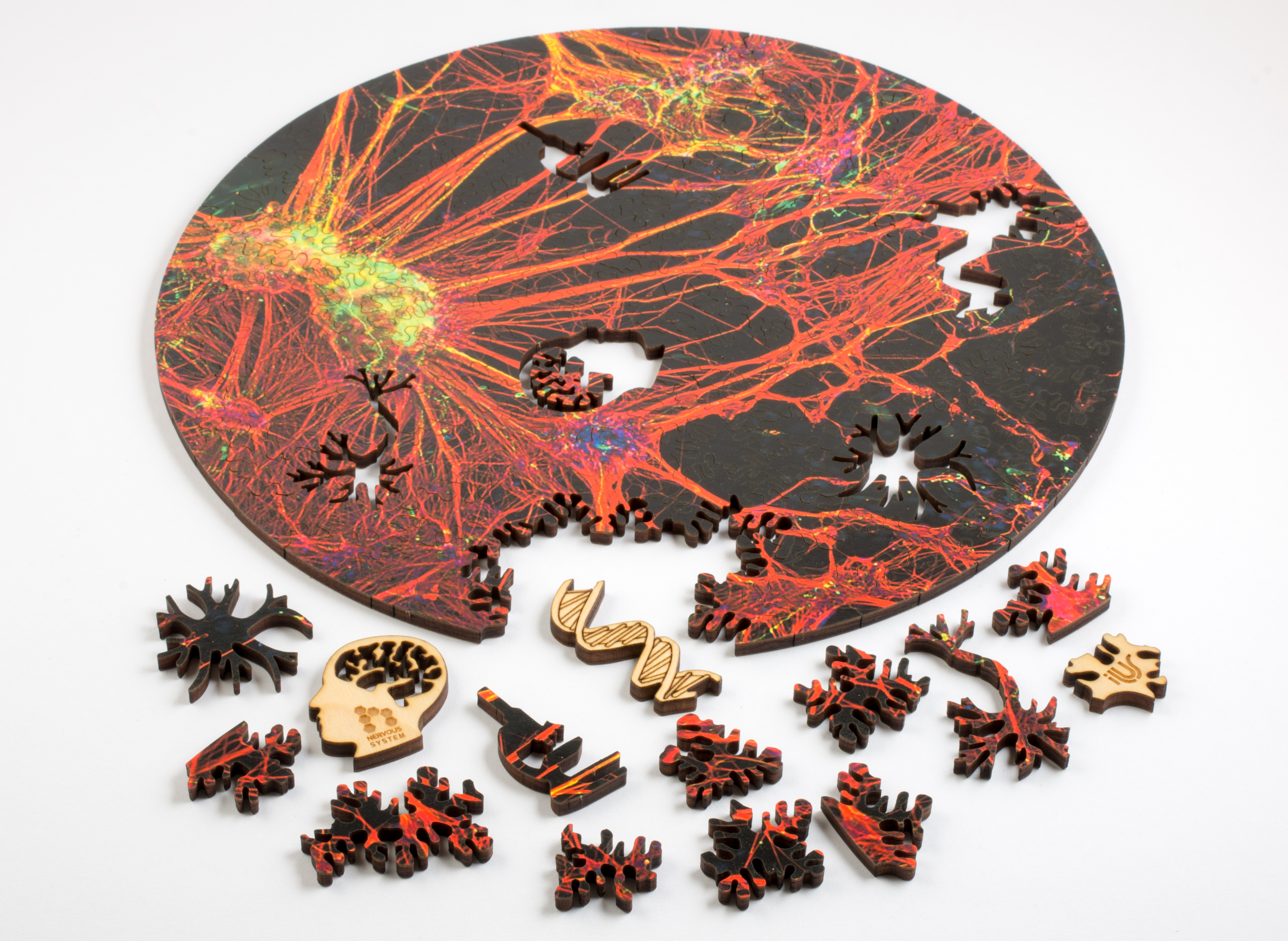
Neuron and Mycoplasma Puzzles with art by David S. Goodsell
Two new biology themed puzzles from Nervous System! They feature the incredible artwork of scientist/artist David S. Goodsell, whose intricate, vibrant watercolor paintings…
Two new biology themed puzzles from Nervous System! They feature the incredible artwork of scientist/artist David S. Goodsell, whose intricate, vibrant watercolor paintings…
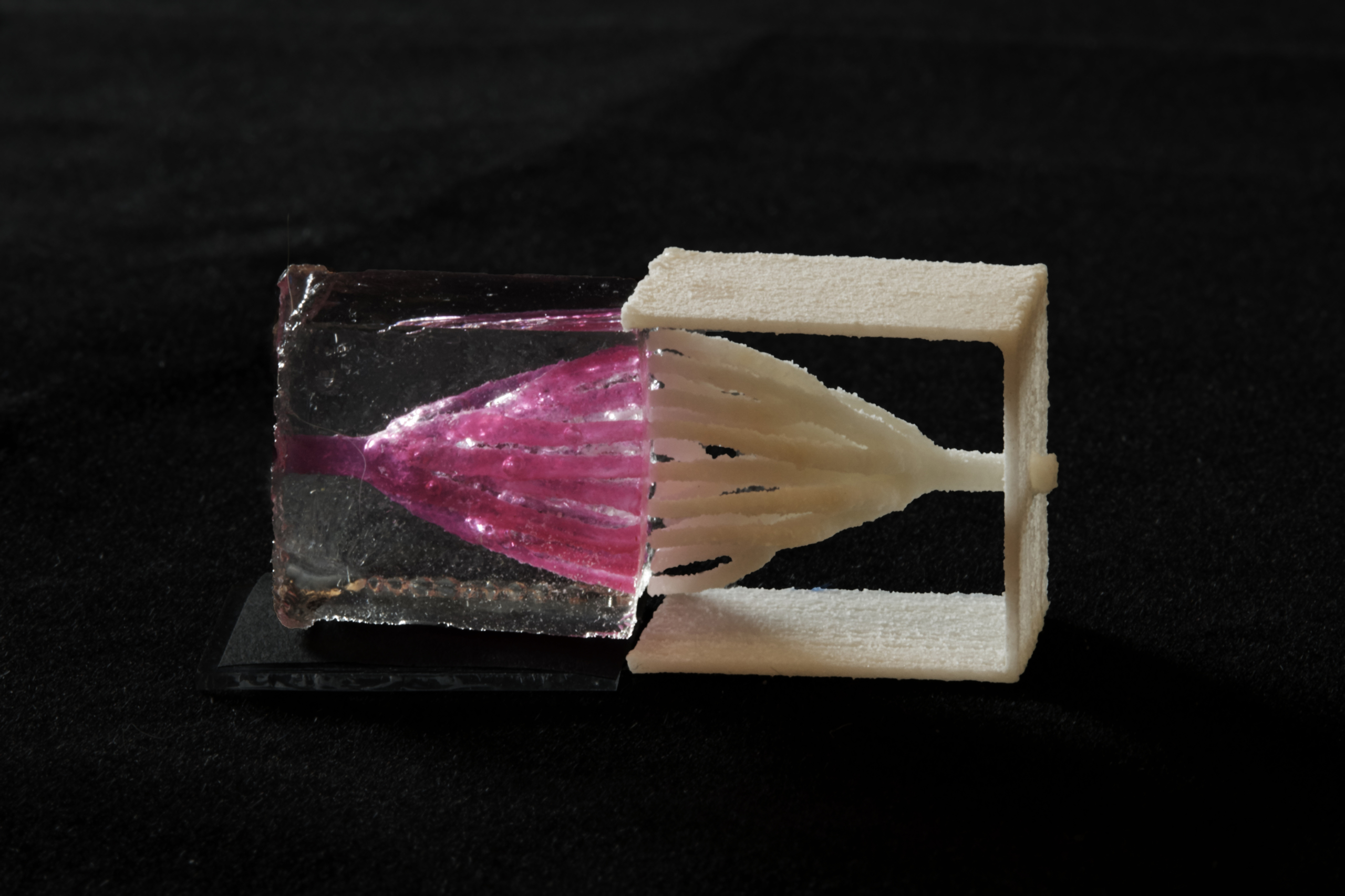
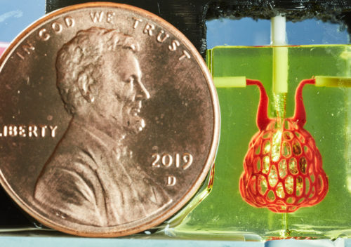
Nervous System collaborated with bioengineers at Rice University to create complex blood vessel networks using generative design and 3D-printed sugar. In a paper…
We founded Nervous System in 2007 with the goal of adapting algorithms and strategies from nature into new ways to design products; but…
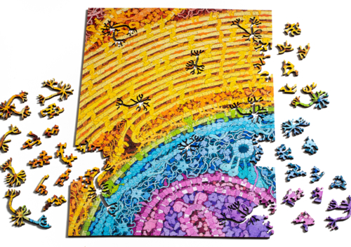
We are excited to announce a new series of generative jigsaw puzzles featuring microscopic art! These laser-cut, computer generated jigsaws feature microscopic photographs…
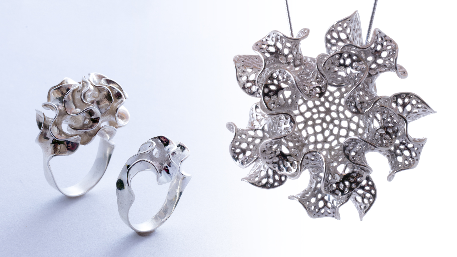
Introducing Floraform, the latest generative design system from Nervous System. Floraform is inspired by the biomechanics of growing leaves and blooming flowers and…
Last weekend we ventured to the Arnold Arboretum to view the blooming lilacs. Although the flowers were spectacular, it was actually a garbage…
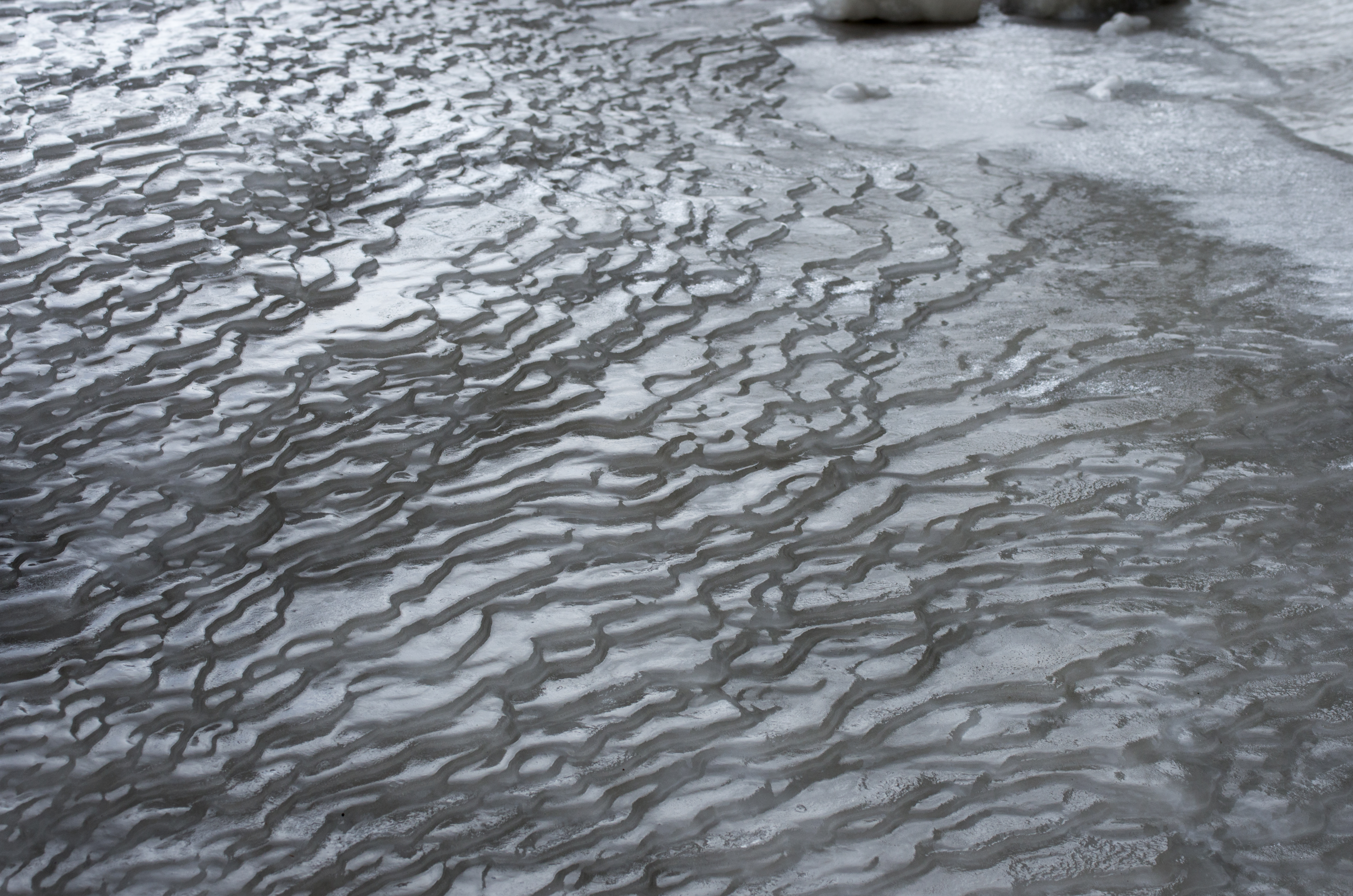
Here in Boston, we’ve been blanketed by a record-breaking 100+ inches of snow. It has been too cold for any of it to…
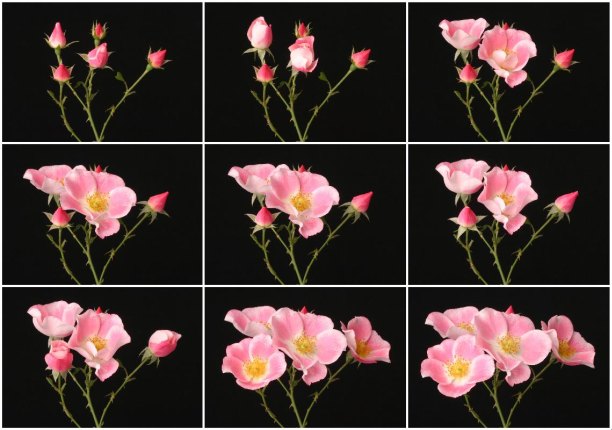
Inside a bud, a flower’s petals lie in wait, a tight bundle of compressed tissue. When the conditions are right, they burst forth,…
I rendered this animation last night of one of my favorite floraform pieces we’ve generated so far. It exhibits an odd, spiraling, 5-form…
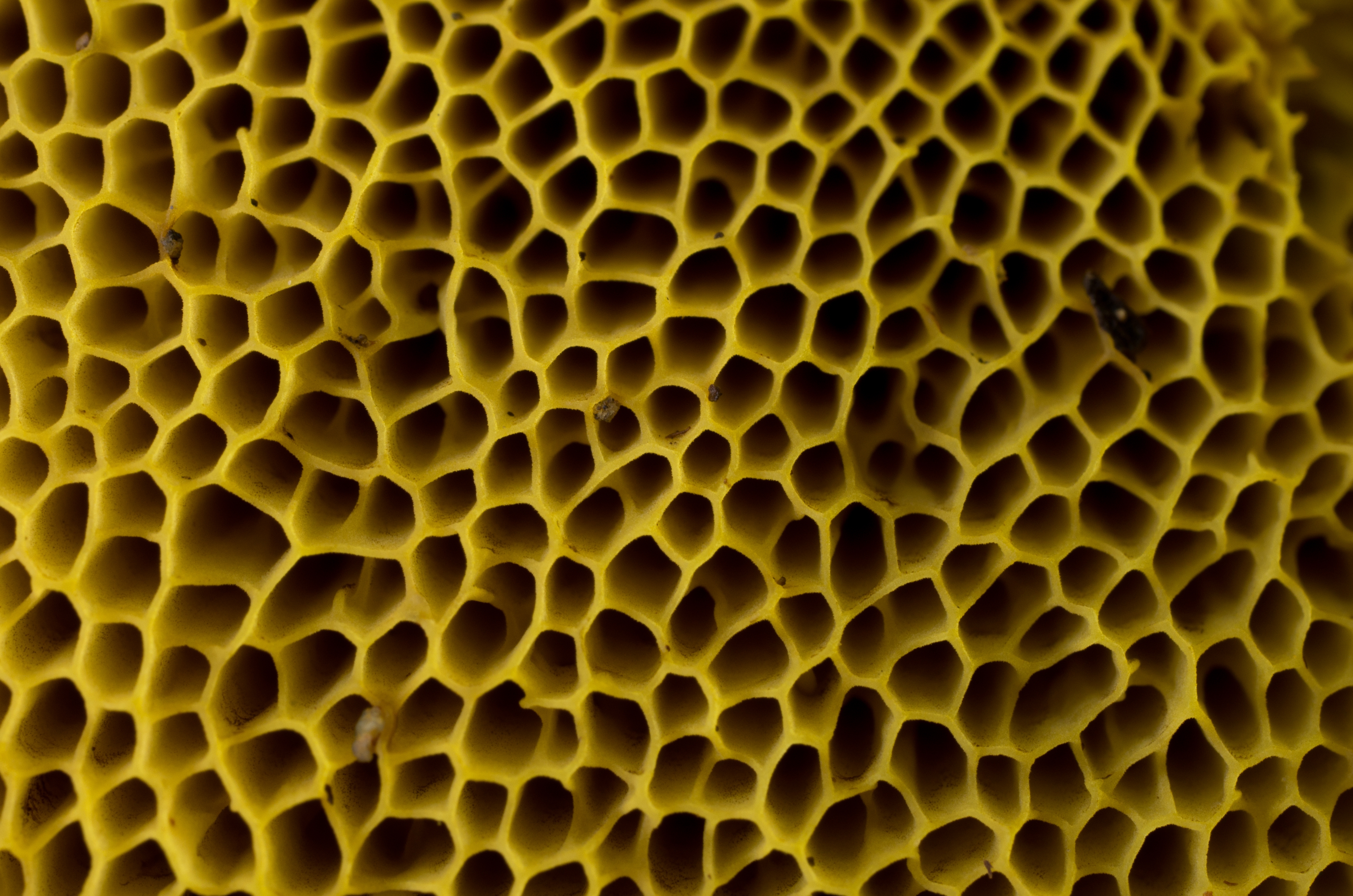
For most, the word fungus evokes images of the gilled mushrooms that appear on our plates or pop up in suburban lawns. But,…